Simulating a mechanistic data driven model of the human spine to predict the growth and spinal curve progression in adolescent idiopathic scoliosis
Grant Recipient: John Sarwak, MD
- Institution:
- Lurie Children's Hospital, Chicago
- Additional Information:
- Final Report
Adolescent Idiopathic scoliosis (AIS) is a structural deformity of the human spine. Both males and females are affected by the disease but the classic case is an adolescent female during puberty. Treatment for AIS includes observation, bracing or surgery. This pilot project aims to understand early AIS induction and progression via 3D modelling of the spine using the patient’s XR and MR images. We expected to gain an understanding of the asymmetrical forces acting upon the spine through this funding. In the future, once the model is validated, we expect that detailed data on these asymmetrical forces will inform non-surgical treatments to aid in curve progression control and if possible, avoid invasive spinal fusion surgery.
Finite element method (FEM) is a powerful tool that helps to understand the functional biomechanics of the spine. Current FEM models of the spine generally account for a single distinct geometry and one set of material properties and are validated with limited experimental cases. This fact raises questions concerning the predictive ability, the range of results and agreement with in vitro and in vivo values of these models.
Our proposed research methodology uses FEM synergistically with bio-mechanical information, image processing, and data science techniques to improve their predictive ability. Initial geometry of the spine was extracted from the x-ray images which are taken from different planes. Later, these images were imported to the FEM software to generate the spine model and perform FE analysis. The x-ray images were provided by Lurie Children’s Hospital. Since the longitudinal growth of long bones and vertebrae occurs in cartilaginous growth plates, in this study the progressive growth will be applied to these plates using a conceptual model corresponding to the biological process of long bone growth. The material properties of different parts will be updated dynamically which is a significant novelty of the proposed methodology. Moreover, research has shown that AIS is associated with low bone mass and supplementing calcium and vitamin D improves bone strength and helps reduce curve progression in AIS. The developed FE model can incorporate the effect of bone mineral density (BMD) on the curve progression as well.
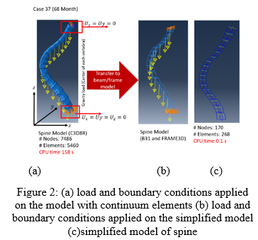
A preliminary result in the first step of the project was a reduced order model. It was developed based on the detailed FE model of the spine. Fig 2 shows the preliminary result of the reduced order model which the CPU time decreases from 158 s to 0.1 s.
The primary outcome measure of success was reached. The primary project was a pilot study with a sample size consisting of 20 patients, 10 control and 10 patients with AIS. To determine the effects of asymmetrical loading forces on the middle column of immature spines in AIS, we:
Aim 1: Used the patient XR and MR images to generate 3D models of the spines using Mimics software for each of the 20 pilot spines.
The student supported through microgrant funding helped in generating these 3D models. Through the funding, he learned research methodology, IS biomechanics, and how to analyze spine XR and MR images.
In the future we will compile the results of our findings and then use the preliminary results of this research to apply for external funding, a NIH grant. We also plan to submit our findings to academic journals for publication. Currently we have a paper under review with the Journal of Applied Mechanics and Engineering.
The funds from the microgrant supported staffing for a medical student intern (Emmett Cleary), to assist in constructing the 3D models during the summer of 2019. Our timeline for the student support was for June 2019-August 2019. A philanthropic grant in combination with support from Northwestern University McCormick School of Engineering supported a research scientist (Ayesha Maqsood) and a PhD mechanical engineering student (Mahsa Tajdari) to produce 3D models of the spine. Materialise Mimics is an image processing software for 3D design and modeling and was utilized to construct the 3D models of the spines from the XR and MR images. The use of the Mimics software and the images was supported by Lurie Children’s Hospital.